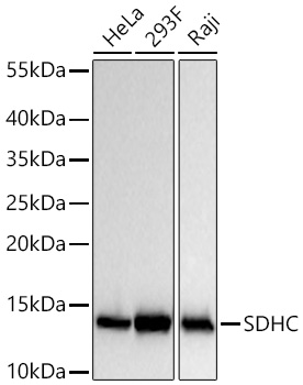
Western blot analysis of various lysates using SDHC Rabbit mAb (A22280) at 1:100000 dilution incubated overnight at 4℃.Secondary antibody: HRP-conjugated Goat anti-Rabbit IgG (H+L) (AS014) at 1:10000 dilution.Lysates/proteins: 25 μg per lane.Blocking buffer: 3% nonfat dry milk in TBST.Detection: ECL Basic Kit (RM00020).Exposure time: 30 s.
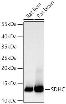
Western blot analysis of various lysates using SDHC Rabbit mAb (A22280) at 1:100000 dilution incubated overnight at 4℃.Secondary antibody: HRP-conjugated Goat anti-Rabbit IgG (H+L) (AS014) at 1:10000 dilution.Lysates/proteins: 25 μg per lane.Blocking buffer: 3% nonfat dry milk in TBST.Detection: ECL Basic Kit (RM00020).Exposure time: 30 s.
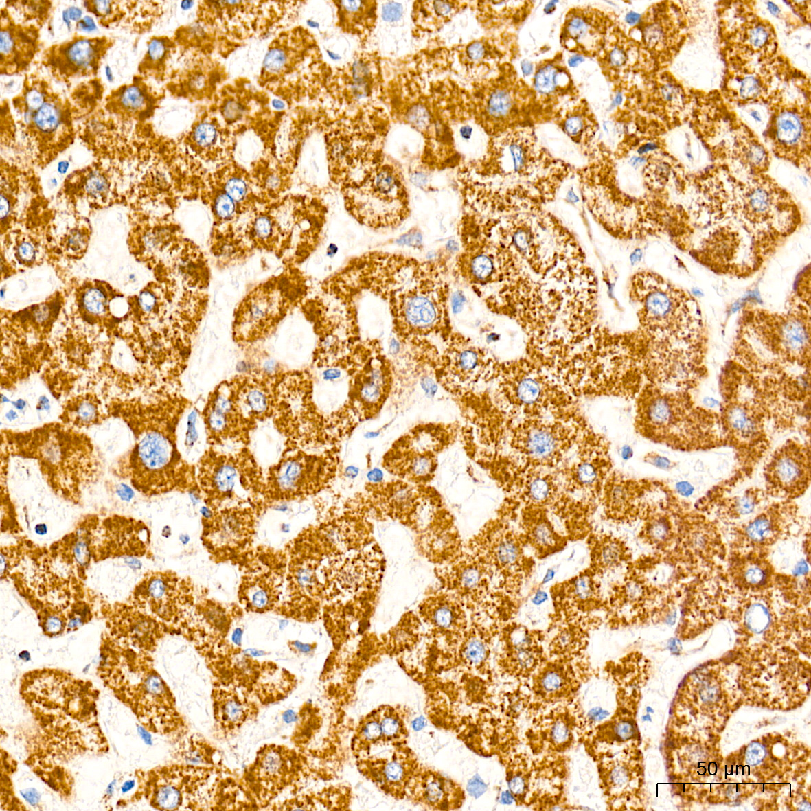
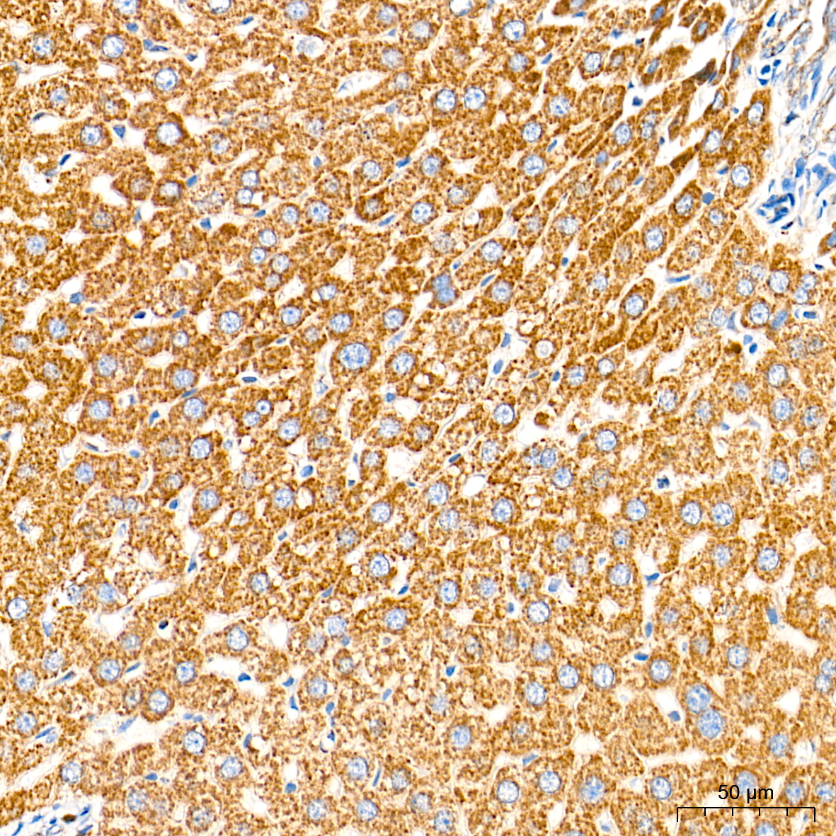
Immunohistochemistry analysis of paraffin-embedded Human liver tissue using SDHC Rabbit mAb (A22280) at a dilution of 1:3000 (40x lens). High pressure antigen retrieval performed with 0.01M Tris-EDTA Buffer (pH 9.0) prior to IHC staining.
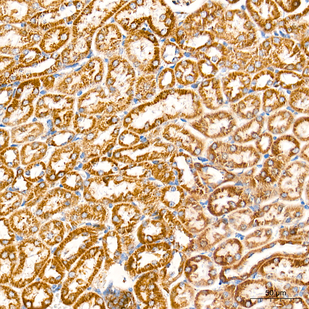
Immunohistochemistry analysis of paraffin-embedded Mouse kidney tissue using SDHC Rabbit mAb (A22280) at a dilution of 1:3000 (40x lens). High pressure antigen retrieval performed with 0.01M Tris-EDTA Buffer (pH 9.0) prior to IHC staining.
Immunohistochemistry analysis of paraffin-embedded Rat liver tissue using SDHC Rabbit mAb (A22280) at a dilution of 1:3000 (40x lens). High pressure antigen retrieval performed with 0.01M Tris-EDTA Buffer (pH 9.0) prior to IHC staining.
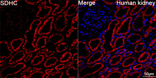
Confocal imaging of paraffin-embedded Human kidney tissue using SDHC Rabbit mAb (A22280, dilution 1:2000) followed by a further incubation with Cy3 Goat Anti-Rabbit IgG (H+L) (AS007, dilution 1:500) (Red). DAPI was used for nuclear staining (Blue). High pressure antigen retrieval performed with 0.01M Citrate Buffer (pH 6.0) prior to IF staining. Objective: 40x.

Confocal imaging of paraffin-embedded Mouse kidney tissue using SDHC Rabbit mAb (A22280, dilution 1:2000) followed by a further incubation with Cy3 Goat Anti-Rabbit IgG (H+L) (AS007, dilution 1:500) (Red). DAPI was used for nuclear staining (Blue). High pressure antigen retrieval performed with 0.01M Citrate Buffer (pH 6.0) prior to IF staining. Objective: 40x.
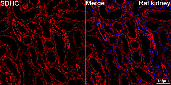
Confocal imaging of paraffin-embedded Rat kidney tissue using SDHC Rabbit mAb (A22280, dilution 1:2000) followed by a further incubation with Cy3 Goat Anti-Rabbit IgG (H+L) (AS007, dilution 1:500) (Red). DAPI was used for nuclear staining (Blue). High pressure antigen retrieval performed with 0.01M Citrate Buffer (pH 6.0) prior to IF staining. Objective: 40x.
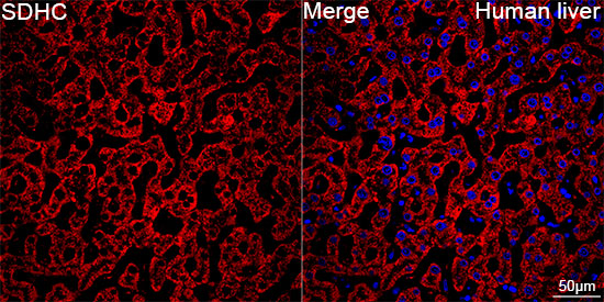
Confocal imaging of paraffin-embedded Human liver tissue using SDHC Rabbit mAb (A22280, dilution 1:2000) followed by a further incubation with Cy3 Goat Anti-Rabbit IgG (H+L) (AS007, dilution 1:500) (Red). DAPI was used for nuclear staining (Blue). High pressure antigen retrieval performed with 0.01M Citrate Buffer (pH 6.0) prior to IF staining. Objective: 40x.
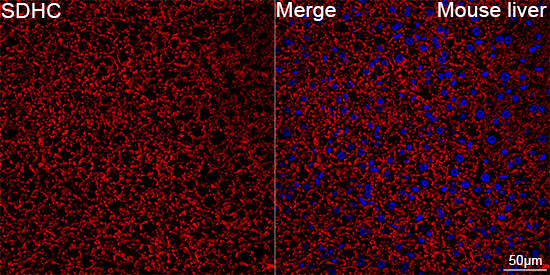
Confocal imaging of paraffin-embedded Mouse liver tissue using SDHC Rabbit mAb (A22280, dilution 1:2000) followed by a further incubation with Cy3 Goat Anti-Rabbit IgG (H+L) (AS007, dilution 1:500) (Red). DAPI was used for nuclear staining (Blue). High pressure antigen retrieval performed with 0.01M Citrate Buffer (pH 6.0) prior to IF staining. Objective: 40x.
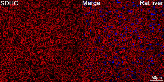
Confocal imaging of paraffin-embedded Rat liver tissue using SDHC Rabbit mAb (A22280, dilution 1:2000) followed by a further incubation with Cy3 Goat Anti-Rabbit IgG (H+L) (AS007, dilution 1:500) (Red). DAPI was used for nuclear staining (Blue). High pressure antigen retrieval performed with 0.01M Citrate Buffer (pH 6.0) prior to IF staining. Objective: 40x.
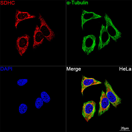
Confocal imaging of HeLa cells using SDHC Rabbit mAb (A22280, dilution 1:2000) followed by a further incubation with Cy3 Goat Anti-Rabbit IgG (H+L) (AS007, dilution 1:500) (Red). The cells were counterstained with α-Tubulin Mouse mAb (AC012, dilution 1:400) followed by incubation with ABflo® 488-conjugated Goat Anti-Mouse IgG (H+L) Ab (AS076, dilution 1:500) (Green). DAPI was used for nuclear staining (Blue). Objective: 100x.
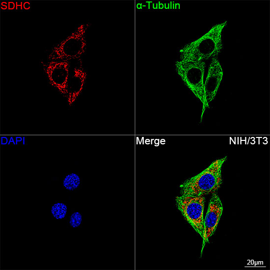
Confocal imaging of NIH/3T3 cells using SDHC Rabbit mAb (A22280, dilution 1:2000) followed by a further incubation with Cy3 Goat Anti-Rabbit IgG (H+L) (AS007, dilution 1:500) (Red). The cells were counterstained with α-Tubulin Mouse mAb (AC012, dilution 1:400) followed by incubation with ABflo® 488-conjugated Goat Anti-Mouse IgG (H+L) Ab (AS076, dilution 1:500) (Green). DAPI was used for nuclear staining (Blue). Objective: 100x.
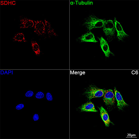
Confocal imaging of C6 cells using SDHC Rabbit mAb (A22280, dilution 1:2000) followed by a further incubation with Cy3 Goat Anti-Rabbit IgG (H+L) (AS007, dilution 1:500) (Red). The cells were counterstained with α-Tubulin Mouse mAb (AC012, dilution 1:400) followed by incubation with ABflo® 488-conjugated Goat Anti-Mouse IgG (H+L) Ab (AS076, dilution 1:500) (Green). DAPI was used for nuclear staining (Blue). Objective: 100x.
请输入产品标签上的lot号,例如4000000001
This gene encodes one of four nuclear-encoded subunits that comprise succinate dehydrogenase, also known as mitochondrial complex II, a key enzyme complex of the tricarboxylic acid cycle and aerobic respiratory chains of mitochondria. The encoded protein is one of two integral membrane proteins that anchor other subunits of the complex, which form the catalytic core, to the inner mitochondrial membrane. There are several related pseudogenes for this gene on different chromosomes. Mutations in this gene have been associated with paragangliomas. Alternatively spliced transcript variants have been described.
首先,一般抗体不推荐客户回收利用,抗体使用之后缓冲体系已经发生改变,不同客户在回收抗体的保存条件上也会有差异,所以抗体回收使用效果无法保证。另外,ABclonal公司也做过一批抗体回收验证测试,测试结果显示不同抗体可回收次数不同,一般效价越高的抗体,可重复使用的次数越多,客户可根据实验情况来确定。
注:我们将孵育完毕后剩余的抗体回收到离心管中置于4℃保存,效价高的抗体可至少保存1周,至少重复利用3次。
武汉爱博泰克生物(ABclonal)科技有限公司是国产品牌,她成立于2011年,公司依托ABclonal美国波士顿抗体与蛋白研发中心、中国光谷生物城(武汉)抗体生产基地以及上海张江分子酶研发中心,凝聚了十余位来自哈佛大学、麻省理工、复旦大学、上海交大、中科院生化细胞所和武汉大学的全球知名分子和免疫学方面博士,组成我们的科学家团队,通过聚焦抗体与酶核心技术,致力于打破国际技术的垄断,将公司打造成为科研工具和诊断原料的国内领导品牌,乃至弯道超越国际巨头。 我们拥有包括兔多克隆抗体、小鼠单克隆抗体、兔单克隆抗体的生产研发平台,同时也有包括WB,IHC,IF,IP,CHIP在内的检测平台,我们对每一支自产的抗体进行了严格的检测。当然,我们部分直销地区也可以帮客户代购进口品牌的产品。同时也有抗体定制服务。ABclonal抗体优势:1,严自检,保质量;2产品多,指标全;3,价格低,货期短。注:ABclonal抗体价格体系详情见附件
ABclonal抗体成分在平时工作当中,常会有客户咨询我们的抗体用的什么buffer进行保存,一般来说,我们的buffer的成分是:PBS含0.03%的proclin300、0.05%牛血清白蛋白、50%甘油;也有一些是PBS含0.03%的proclin300,50%甘油。防腐剂 Proclin 300活性成分主要是2-甲基-4-异噻唑啉-3-酮(MCI)和5-氯-2-甲基-4-异噻唑啉-3-酮(CMCI)。ProClin生物灭活剂能够迅速穿透细胞膜,抑制对细胞呼吸至关重要的特定酶,因此一接触微生物有机体就会立即抑制细胞活性。ProClin的多个特定毒性位点可以防止微生物产生高水平的耐药性。